


Hasanain Kambari and Jayden Dixon
Millions of Americans with diabetes (approximately 1 in 5) face the risk of eventually going blind due to: diabetic retinopathy, a condition that affects blood vessels in the retina, the light-sensitive tissue at the back of the eye. The condition is difficult to detect in the early stages because many people do not show symptoms right away (although symptoms do not appear right away). 2021 survey We have identified important biomarkers that may one day aid in early detection). In later stages, the damage is often irreversible.
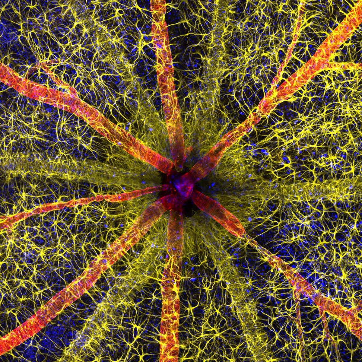
Hassanain Kambari’s research at the Lions Eye Institute in Perth, Australia, focuses on early detection of diabetic retinopathy and the possibility of reversal, including taking precise images of the tiny micron-sized blood vessels in the eye. I’m guessing. With the help of his colleague Jayden Dixon, he created a winning image for 2023. Nikon Small World Microphotography Competitiona detailed depiction of the rodent optic disc.
The annual contest, now in its 49th year, is designed to highlight “amazing images from scientists, artists, and micrographers from around the world, reflecting all their experiences and backgrounds,” according to Nikon. “I’m always in awe,” communications manager Eric Flem said, adding. Let’s consider how these advances make it possible to create art from science for the general public to enjoy. ” microscopic photography This involves attaching a camera to a microscope (either a light microscope or an electron microscope) so that the user can take pictures of objects at very high resolution.british physiologist richard hill norris He was one of the first to use this technique to study blood cells in 1850, and from the 1970s onwards the technique became increasingly recognized as an art.
Kambari used several techniques to capture the winning images, including achieving high enough resolution to display microscopic blood vessels as well as developing protocols to color code different cell types. challenges had to be overcome. Astrocytes are shown in yellow, contractile proteins in red, and retinal vasculature in green.
“The visual system is a complex and highly specialized organ, and even relatively small disruptions to the retinal circulation can cause catastrophic vision loss,” Kambali said. “I entered this competition as a way to introduce the intricacies of the retinal microcirculation.A competition like this not only celebrates the hard work and passion of the participants, but also attracts young scientists and encourages them to pursue a career in STEM. It could also inspire me to pursue that. It certainly inspired me.”
Here are the remaining top 20 entries from this year’s contest. They range from stunning images of igniting matchsticks and extreme close-ups of tarantula fangs to detailed peeks at crystallized sugar syrup and the scales of a Chinese moon moth’s wing.You can check out complete list of winnersas well as a few honorable mentions. here.
-
2nd place: A matchstick that ignites on the friction surface of a matchbox.
ole bierfeld
-
3rd place: Breast cancer cells.
Malgorzata Lisowska
-
The poisonous fangs of a small tarantula.
john oliver dam
-
Autofluorescent protective hairs that cover the leaf surface Eleagnus augustofolia Exposure to ultraviolet light.
david maidland
-
A slime mold whose capillary fibers are visible through a translucent peritoneum.
timothy boomer
-
Mouse embryo.
Grigory Timin and Michael Milinkovich
-
Caffeine crystals.
Stefan Eberhardt
-
Cytoskeleton of a dividing myoblast: tubulin (cyan), F-actin (orange), nucleus (magenta).
Vaibhav Deshmukh
-
Motor neurons are grown in a microfluidic device to separate the cell body (top) and axon (bottom). Microtubules are green and growth cones are red.
Melinda Beccari and Don W. Cleveland
-
Crystallized sugar syrup.
Diego Garcia
-
A cuckoo bee standing on a flower.
Sheriff Abdallah Ahmed
-
Blood and lymphatic structure of adult mouse ear skin
Satu Pavonsalo and Sinem Karaman
-
Sunflower pollen on an acupuncture needle.
john oliver dam
-
fluorescence image of Acropora sp. shows symbiotic zooxanthellae and individual polyps.
Pichaya Lertvilai
-
carbon nanotube.
Diego Garcia
-
Chinese moon moth wing scales.
Yuanji
-
A cryptocrystalline micrometeorite placed on a test sieve.
scott peterson
-
Stomata in the epidermis of a peace lily leaf.
Marek Misch
-
Head of an adult transgenic zebrafish showing blood vessels (blue), lymph vessels (yellow), skin and scales (magenta).
Daniel Castranova and Brant Weinstein


