


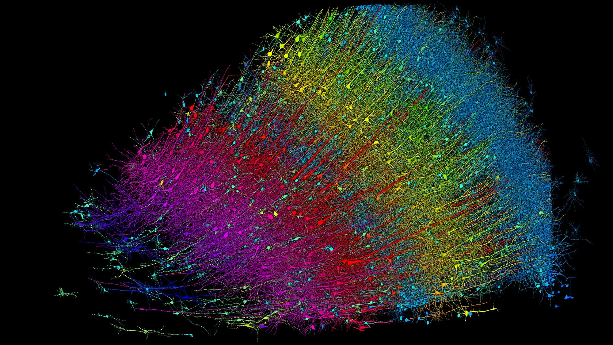
One cubic millimeter is small no matter how you look at it. It is hardly noticeable – specks, specks or crumbs. However, if we look closely, we can see the entire world within the particles of matter. A team of neuroscientists and engineers, aided by machine learning tools, charted a cubic millimeter volume of the human brain at nanoscale resolution, tracing every neuron, synapse, blood vessel, and supporting cell within the fragment. and reconstructed his 3D model of the tissue. . Although it is only a millionth of the brain’s total volume, it is the most detailed map of a portion of human brain matter ever created. It could spark a wave of scientific discoveries about neurological diseases, the structure of the brain, and the origins of our behavior.
“In some respects, our dataset is very small.” Jeff Rismansaid the co-senior investigator, neuroscientist and professor of molecular and cell biology at Harvard University. pop science. “But when you go inside, it doesn’t feel small because it’s like a giant forest. It’s a very small forest, but it’s a very, very, very complex forest,” he added.
All of its complexity is revealed in studies documenting the construction of this comprehensive brain map or “connectome.” Released on May 9th in diary science. The first connectome was from the C. elegans brain. Completed in 1986. Since then, neuroscientists have continued to draw larger and more complex pictures of brains, including those of fruit flies, maggots, tadpoles, and earthworms. However, the human brain presents unique challenges for mapping in that it is complex and difficult to access. The new partial human connectome is available online For anyone to explore.
“This is not only an incredible technological feat, but also a tool and resource that is truly meant to be shared with the world and get all of our scientific information out there.” Tim Moscasays a neuroscientist at Thomas Jefferson University who was not involved in the new study. pop science. “This group has done a great job of designing all the new tools and pipelines and making them available to anyone who wants to look at this, think about it, or use it for research.”

serve brain pizza
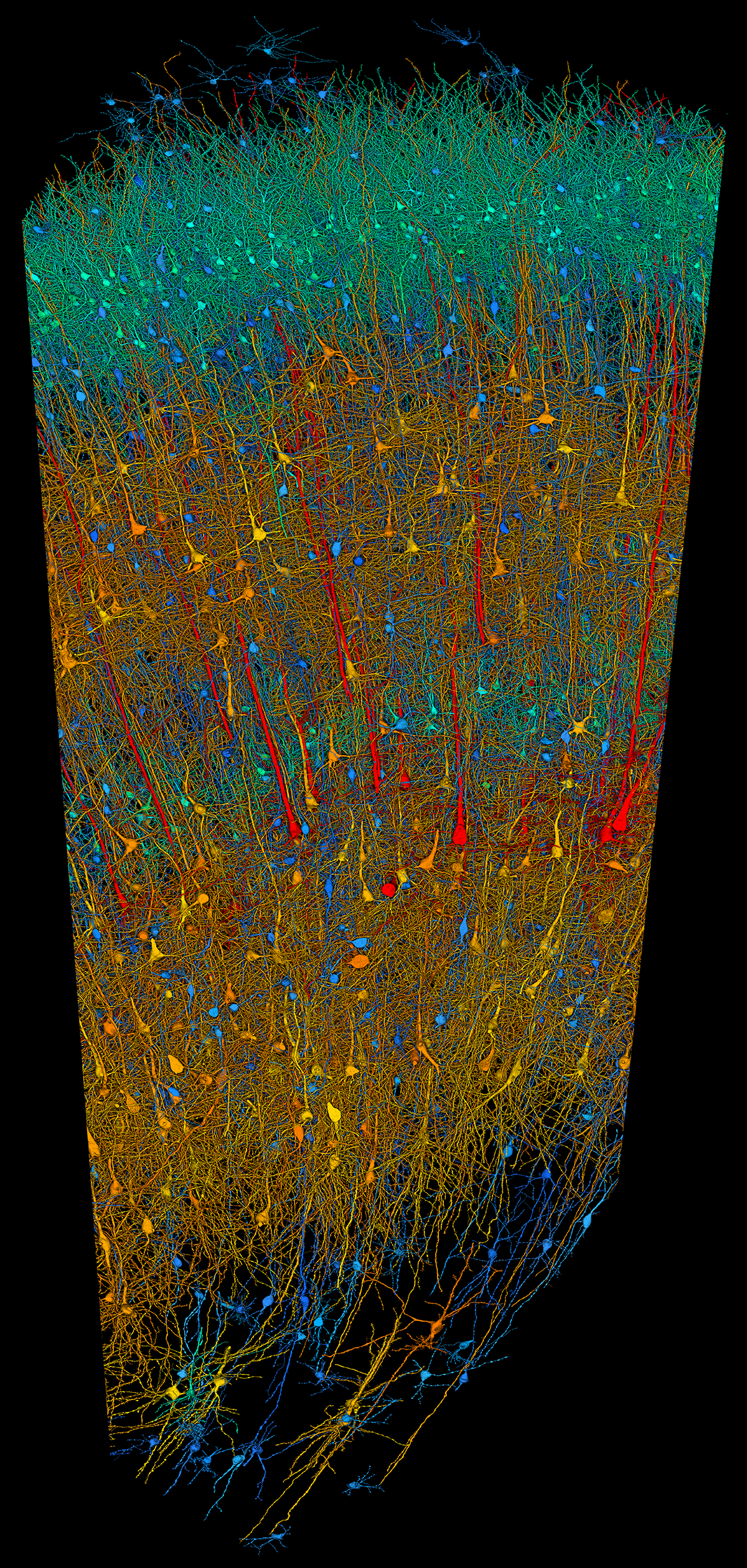
Study samples were collected from anonymous patients who underwent epilepsy surgery more than 10 years ago. Surgeons removed a small piece of the temporal lobe to access and treat the underlying lesion, immediately saving the tissue and later sharing it with scientists. The total volume of the debris is approximately 1 cubic millimeter, but it is not cubic in shape. Rather, “it’s like a thick pizza, but not as thick,” Lichtman says. This blunt triangular mass, which is longer than it is wide, allowed the researchers to capture parts of all six layers of the cerebral cortex, which are 3 mm thick.

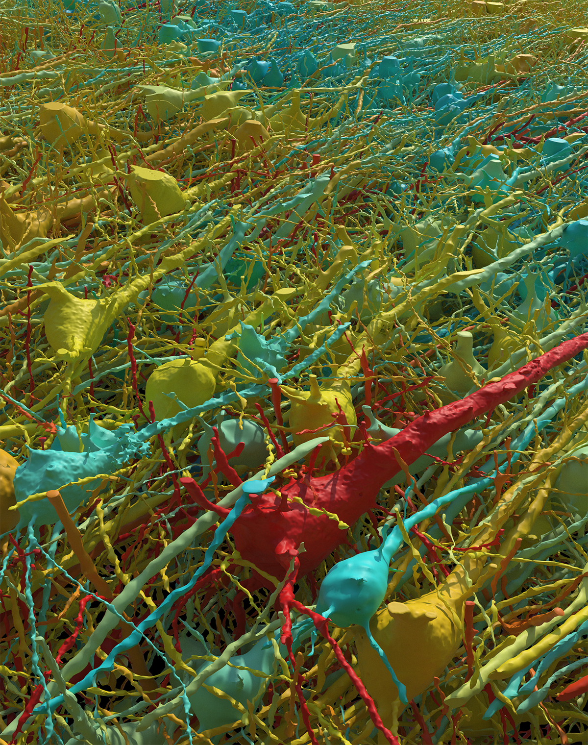
The first step to mapping the brain pizza is Slice it It is divided into 5,019 individual cross-sections (each 30 nanometers thin) that are attached to the tape using a specially designed machine that cuts with a diamond knife. From there, the researchers spent a full year carefully imaging each slice with an electron microscope. The slices were then digitally aligned and stitched together, and using multiple machine learning tools he filled out a 3D form, labeling and coloring each component.
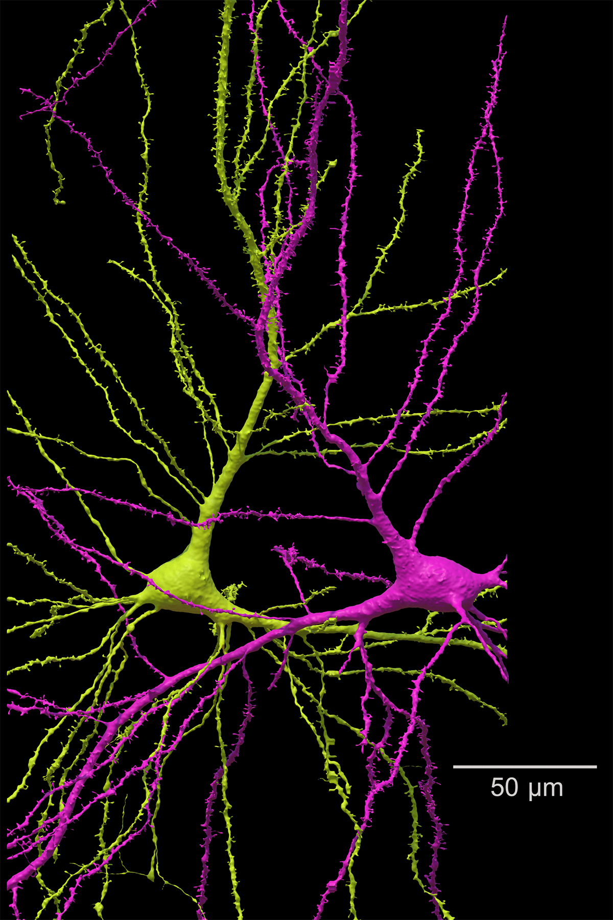
The study found that the neuron density in this region was 16,000 neurons per cubic millimeter, about one-third lower than previous estimates for the same brain sections and 10 times lower than corresponding sections of mouse brains. 1/2 lower. Glial cells, the glue that holds brain tissue together, outnumber neurons in the fragment by a two-to-one ratio.
neural explorer
Although the physical size of brain fragments may be very small, the level of detail means that the data collected by the mapping effort is enormous. The reconstructed segment has a digital size of 1.4 petabytes, or 1,400 terabytes (equivalent to the storage capacity of about 2,800 average laptops). There are many things that could be discovered, such as individual neural circuits, previously unobserved cell ratios and shapes, and the composition of each cortical layer.
“It’s like an explorer landing on a new island,” says Lichtman. “If you keep looking around, you keep finding new things.”
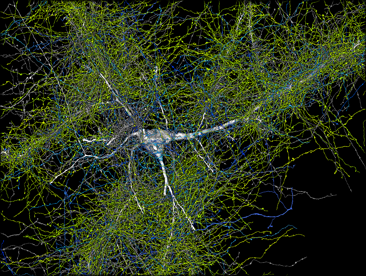
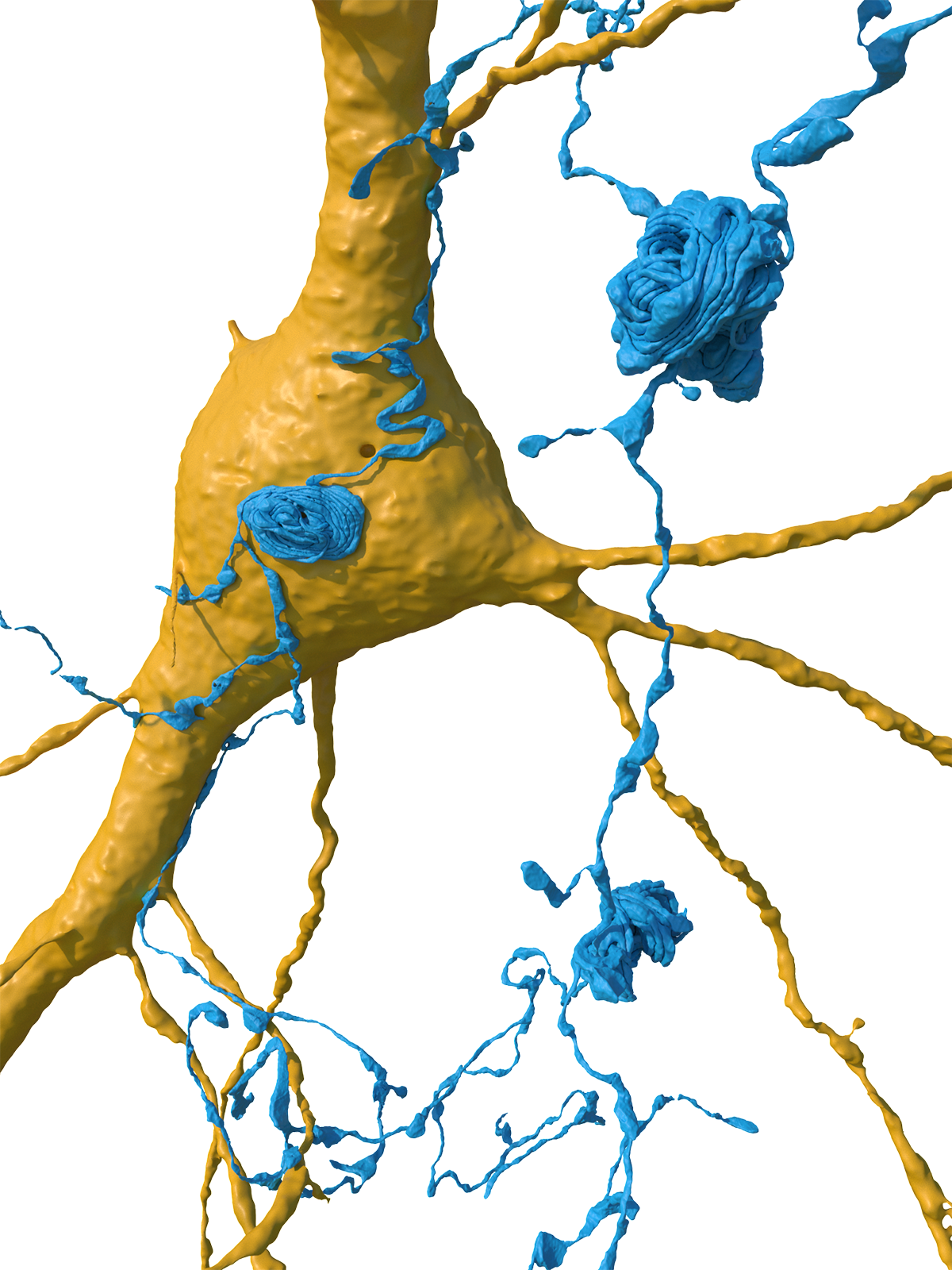
Already, Lichtman and his many collaborators have made some interesting observations. Among the roughly 150 million synapses they mapped, they found a rare type of particularly strong connection. In most cases (96.5%), the axon (the neuron’s outgoing transmission line) formed its single connection with the target cell. Some (about 3%) he made two connections. However, less than 0.01% of them formed four or more synapses, including some axons and target cells connected at more than 50 points.

“There’s always been a theory that there are sort of super connections between certain cells,” Mosca says. “But it’s something we never had the determination to prove…Now we know it exists and can pursue the question of what it does.” Lichtman’s current The hypothesis is that these highly strengthened connections are a kind of superfast pathway that enables “automatic use of the brain” for established and learned behaviors.
Another new observation: many dendrites (branching extensions of neurons that typically receive input) appear to be mirror images of each other, allowing them to be arranged in one of two directions out of an infinite number of three-dimensional possibilities. pointing symmetrically. “We had never seen anything like it. [before]” says Lichtman. “Why would they do that? I don’t understand… [it is] It’s a complete mystery. ”

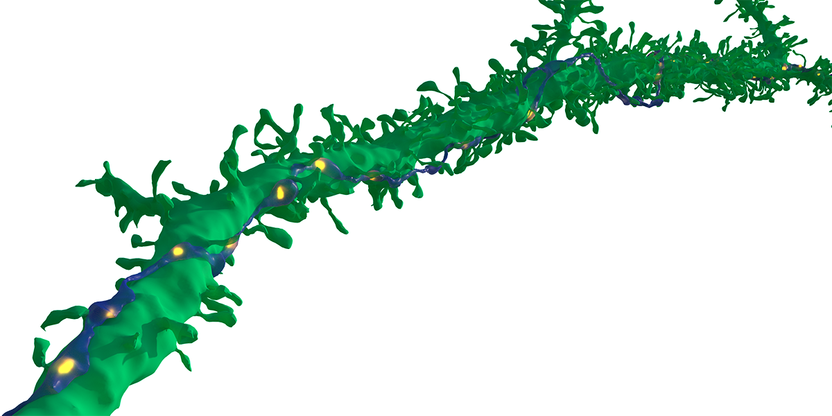
The scientists also discovered a new type of unexplained structure, which they termed an “axonal whorl,” which appears to be a tangle of long axonal cables. Although not all neurons, some axons contained multiple knots. biren jainismco-senior study author and senior staff scientist at Google, leads the company’s research. connectomics Research team. Again, the function and cause of these whorls are unknown. “We didn’t expect to find a structure like this. It’s very strange… like a big hodgepodge of wiring that defeats the purpose of wires in the first place, which is to go from one place to another. It’s something.”

These three discoveries are likely just the tip of the iceberg. “The data set is so large that no single person or research group can study it.” [all]But many people can do it,” Lichtman says. Due to the open nature of the project, 200 papers Jain points out that he has been talking about brain restructuring since it was first published as a preprint.
In addition to being a fundamental breakthrough in science, discoveries from this partial connectome could ultimately help us better understand and treat brain diseases. “Being able to measure the neural wiring of the human brain in such detail opens up incredible opportunities to promote human health,” he says. andrew leifer, a physicist and neuroscientist at Princeton University, was not involved in the project. “You can also imagine comparing different brains to understand how the brain’s wiring changes when a healthy brain becomes diseased or malfunctions.” he added.
Challenging the future frontier
But while there is much to discover, there are also limits. The automated machine learning methods that were key to making such a large-scale effort possible contain errors that require human oversight to correct. Editing is an ongoing project, a community science effort that anyone can edit if they wish. Apply to participate.
The sample is also just a small portion of one person’s brain. There are still many things we can’t infer about the human brain in general, or other brain regions beyond the temporal lobe based on this single fragment, without more samples and comparative maps, Lichtman said. points out.
And perhaps most importantly, this part of the brain came from a person who had undergone surgery for epilepsy. This may not represent a “normal” brain, and there’s no way to know for sure until more parts are evaluated, Jain and Lichtman say. “But we teeth We have a lot of follow-up planned on this,” Jain added.

The team has ambitions to construct multiple partial connectomes representing additional human brain samples. They are also working on the zebrafish connectome and plan to work on increasingly larger parts of the mouse brain. Because mammalian brains share many similarities, the complete mouse connectome could provide new insights not only into our own brains but also into the evolution of brains across animals. Lichtman says.
At this point, Lichtman says, with currently available technology (and ethical implications), Brian’s complete connectome in humans is “a bridge too far.” “We’re literally a million times further away from that,” Jayne says. But through this work, scientists have taken an early (albeit small) step in that direction, and even the tiniest peephole can become a gateway to an entire universe of knowledge.
“I want people to think about this the same way they think about the Hubble telescope or the James Webb telescope,” Lichtman says. “We are peering into unknown territory, but it is an area that is far more relevant to us than the distant universe. It is this inner space that each of us carries on our shoulders and utilizes. But we know almost nothing about it.”


